Zinc ion flux during mammalian sperm capacitation
Kerns et al., 2018 · Nature Communications
Single Cell Phenomics Center
High-throughput microscopy meets flow cytometry and artificial intelligence—opening a new window into cellular heterogeneity.
Explore capabilities ↓
Overview
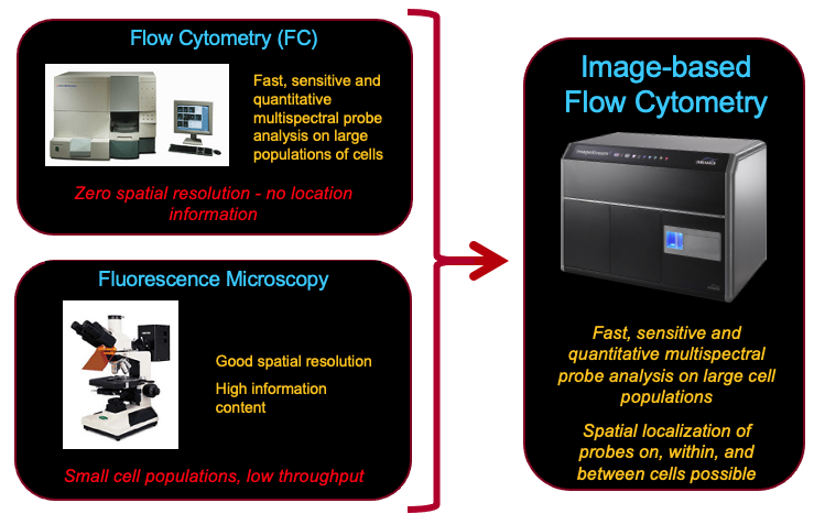
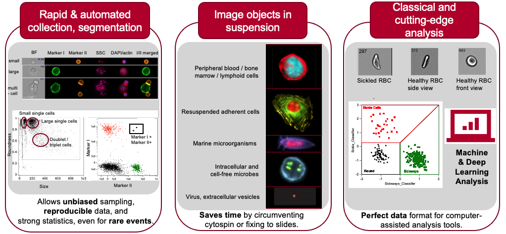
The Single Cell Phenomics Center brings advanced image-based flow cytometry to Iowa State researchers and external collaborators. The technology works like a high-throughput microscope, imaging thousands of cells in seconds while collecting brightfield, side scatter, and fluorescence data.
 01 · The platform
Two strengths, one instrument
Quantitative flow data is paired with detailed cellular images in a single platform. Samples can be viewed in brightfield, side scatter, and across a wide fluorescent palette—with extended depth of field when the research question calls for it.
Research applications
Artificial intelligence
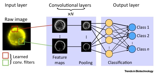
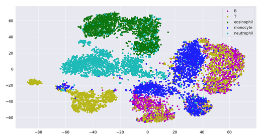
Image outputs integrate naturally with machine- and deep-learning approaches, enabling label-free classification and richer phenotypic discovery.
Selected publications
Kerns et al., 2018 · Nature Communications
Zigo et al., 2019 · Scientific Reports
Kerns et al., 2020 · Antioxidants
Kerns et al., 2020 · IJMS
Internal & external access
Services are available to Iowa State principal investigator labs and external stakeholders. Equipment scheduling and billing are managed through iLab.
Schedule through iLab ↗Project inquiry
Tell us about the cells or particles, approximate dimensions, available labels, desired measurements, sample count, and timeline. We can help assess fit before you schedule through iLab.
Submitting prepares an email in your own mail application; no project information is stored on this website.