⌁
Multi-omics
Proteomics, lipidomics, metabolomics, genomics, and epigenomics reveal the systems behind reproductive success.
Research narrative →Molecular reproductive physiology
We investigate the cellular and molecular mechanisms that shape livestock sire fertility—turning single-cell insight into tools for a more productive, sustainable future.
From a single cell
to a healthier herd.
At the intersection of biology, data, and animal agriculture, we’re building a clearer picture of male fertility—one cell at a time.
Four integrated programs move from molecular discovery to precision diagnostics.
Explore ↗02Peer-reviewed scholarship with corresponding- and first-author work emphasized.
View record ↗03Invited talks, abstracts, posters, scientific meetings, and international instruction.
Explore exchange ↗04Anatomy, physiology, entrepreneurship, AI-enabled learning, and mentorship.
Explore ↗Our research
Our multi-omic approach connects molecular signatures with reproductive outcomes in boars, bulls, and other sires.
Proteomics, lipidomics, metabolomics, genomics, and epigenomics reveal the systems behind reproductive success.
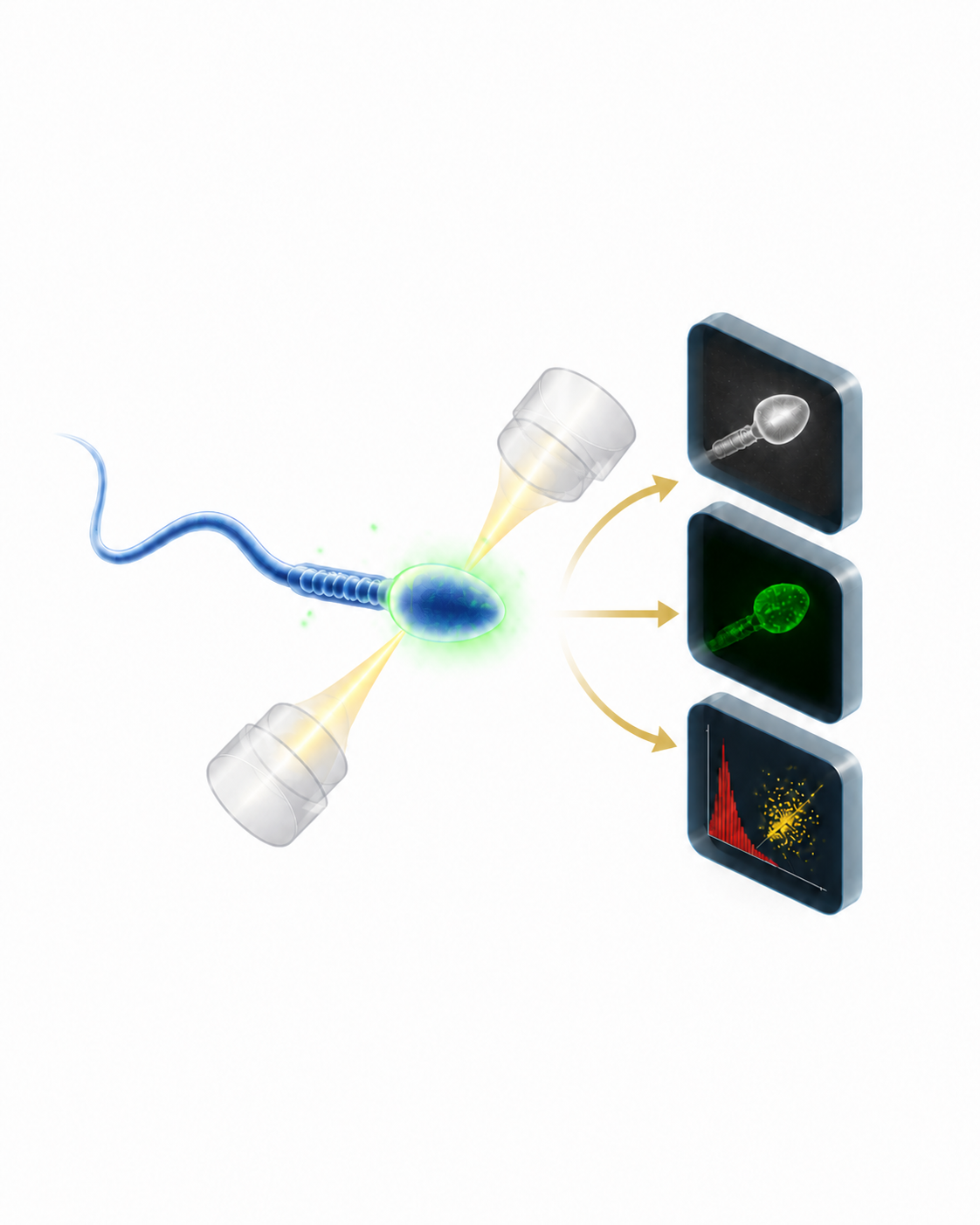
Research narrative →High-throughput cellular imaging paired with artificial intelligence identifies meaningful fertility biomarkers.
Visit the center →Advanced assays turn discovery into practical insight for producers, breeders, and industry partners.
Andrology Analytics →Why it matters
Improving our ability to predict and understand sire fertility can strengthen livestock efficiency while reducing the resources needed to produce food.
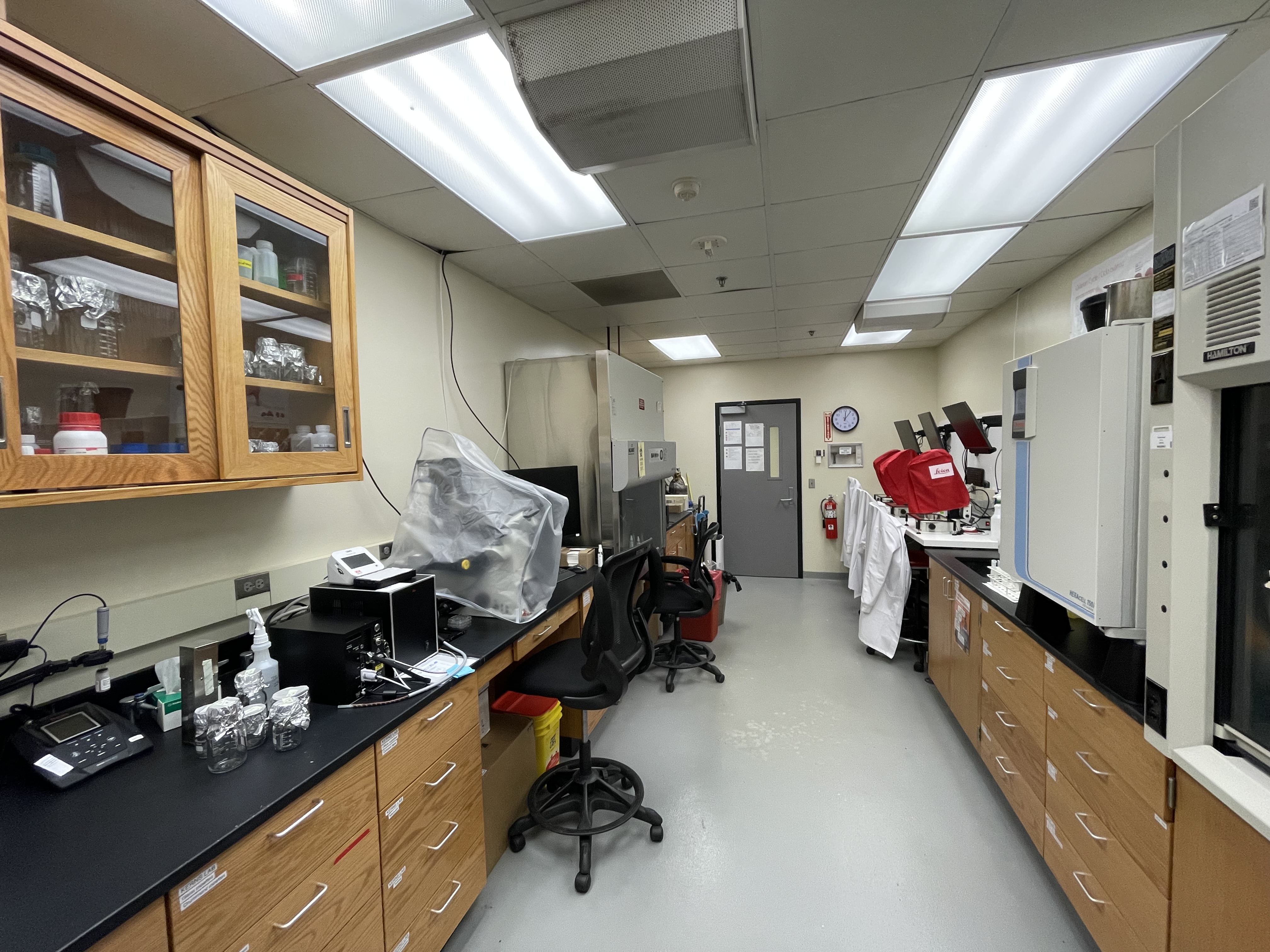
Read the Iowa State storyBuilt for discovery
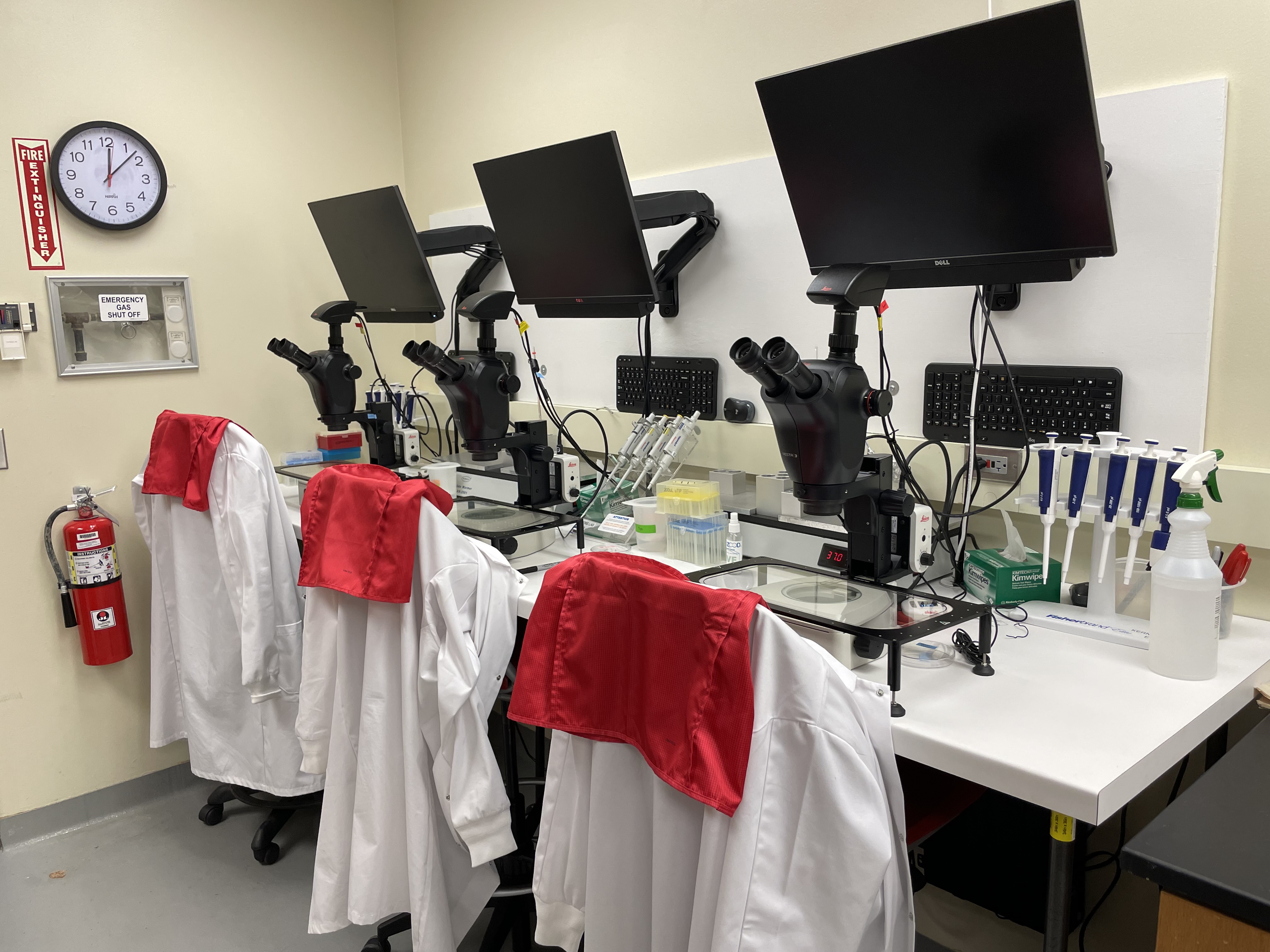
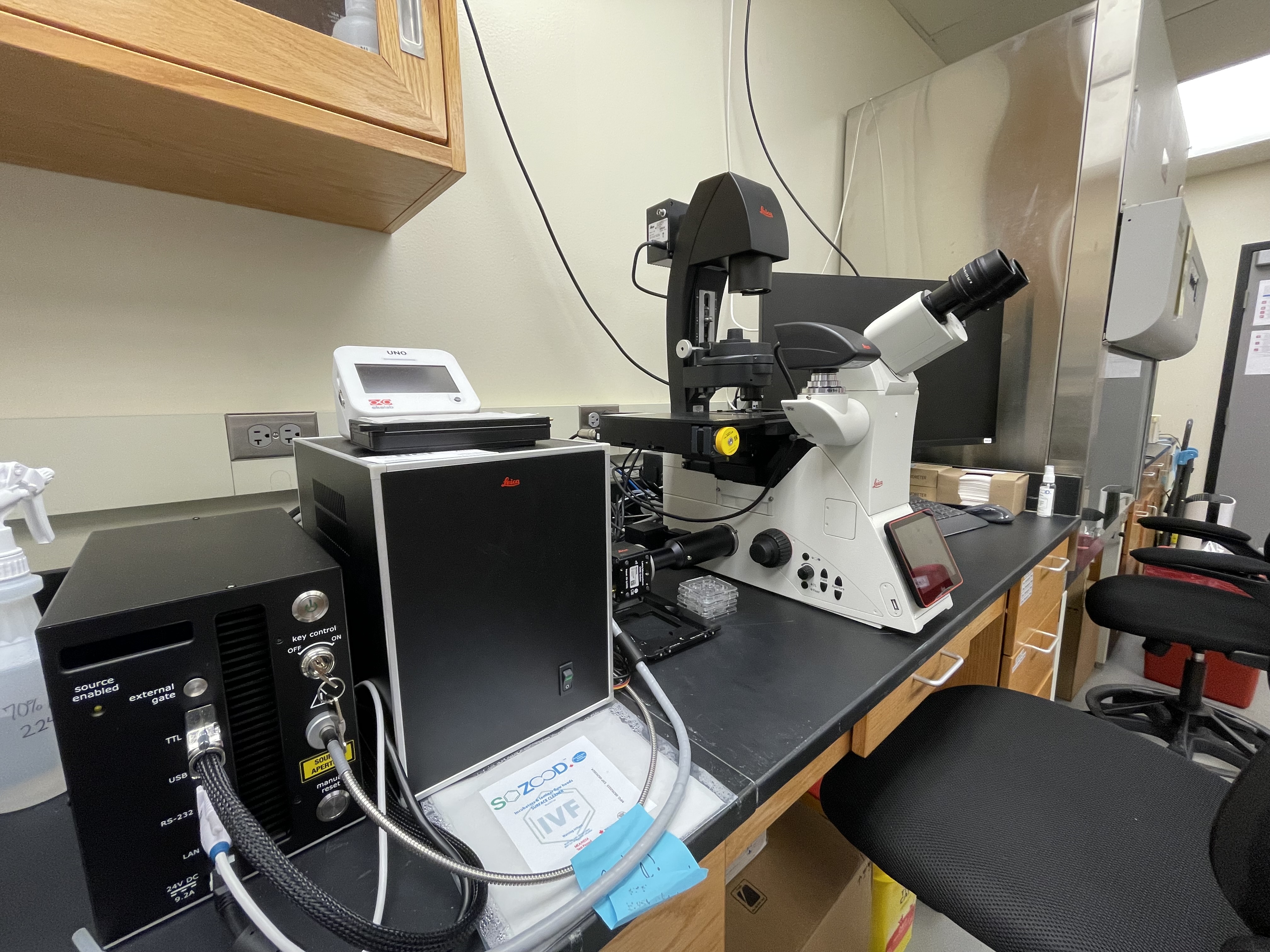
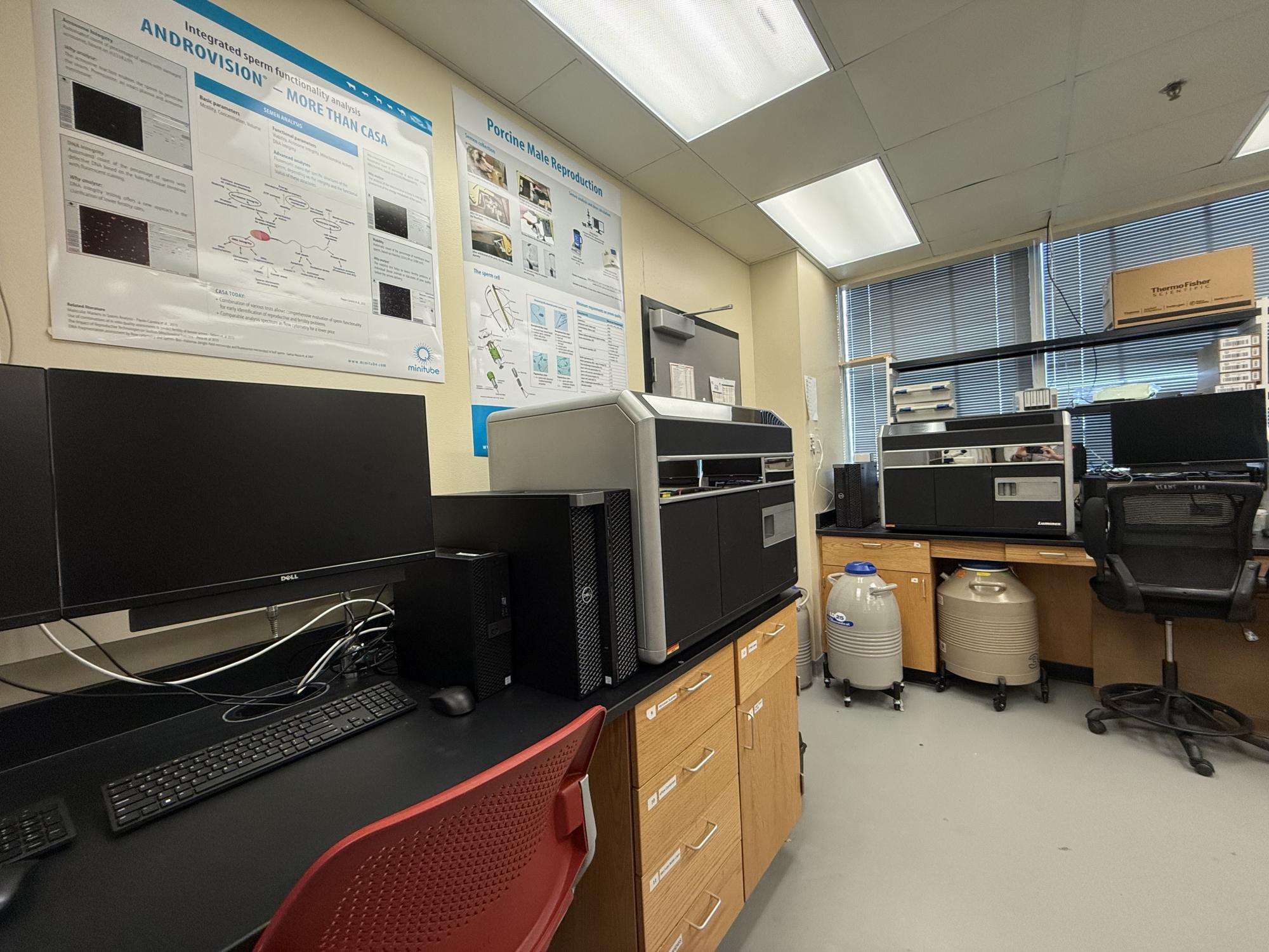
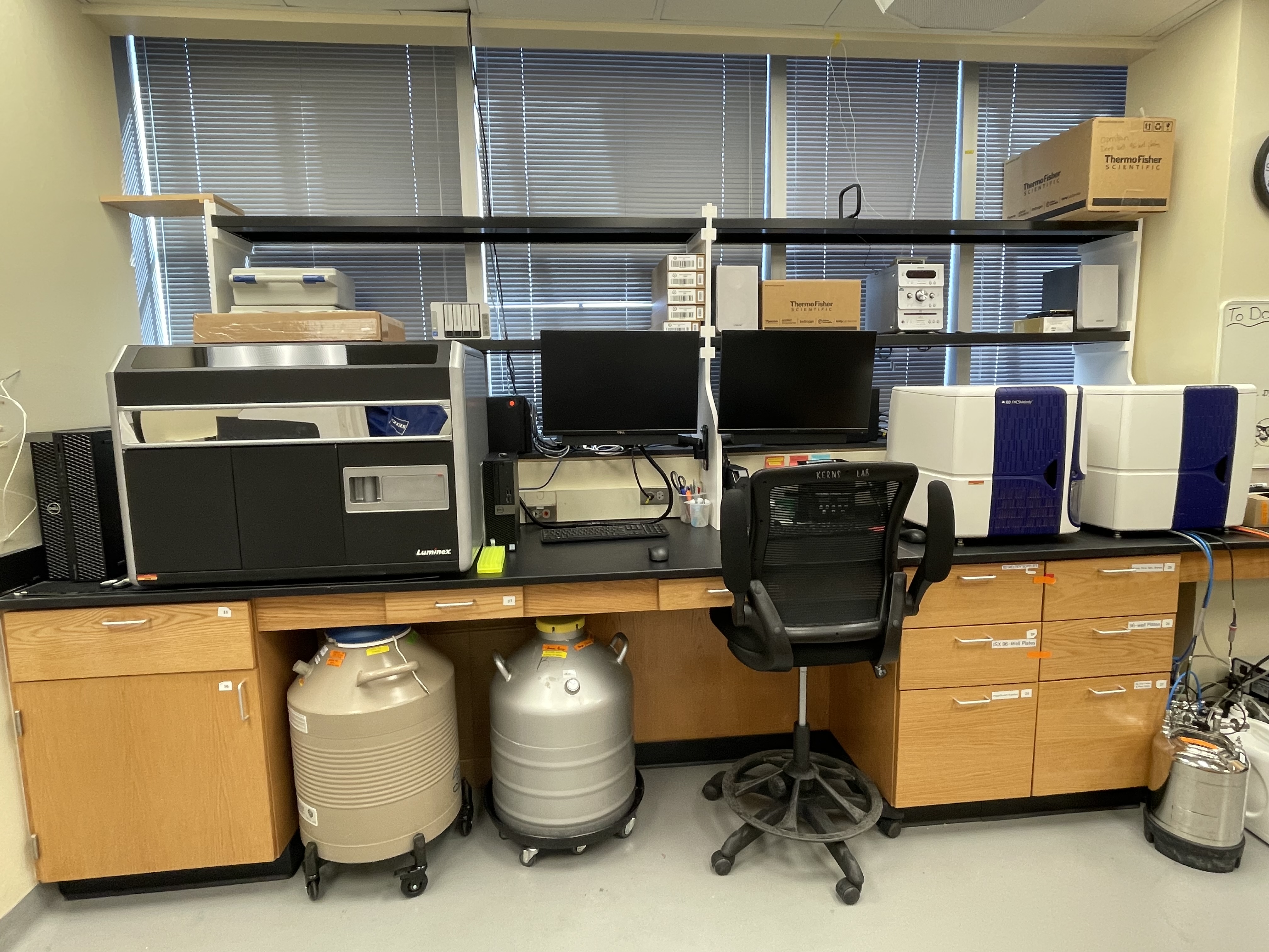
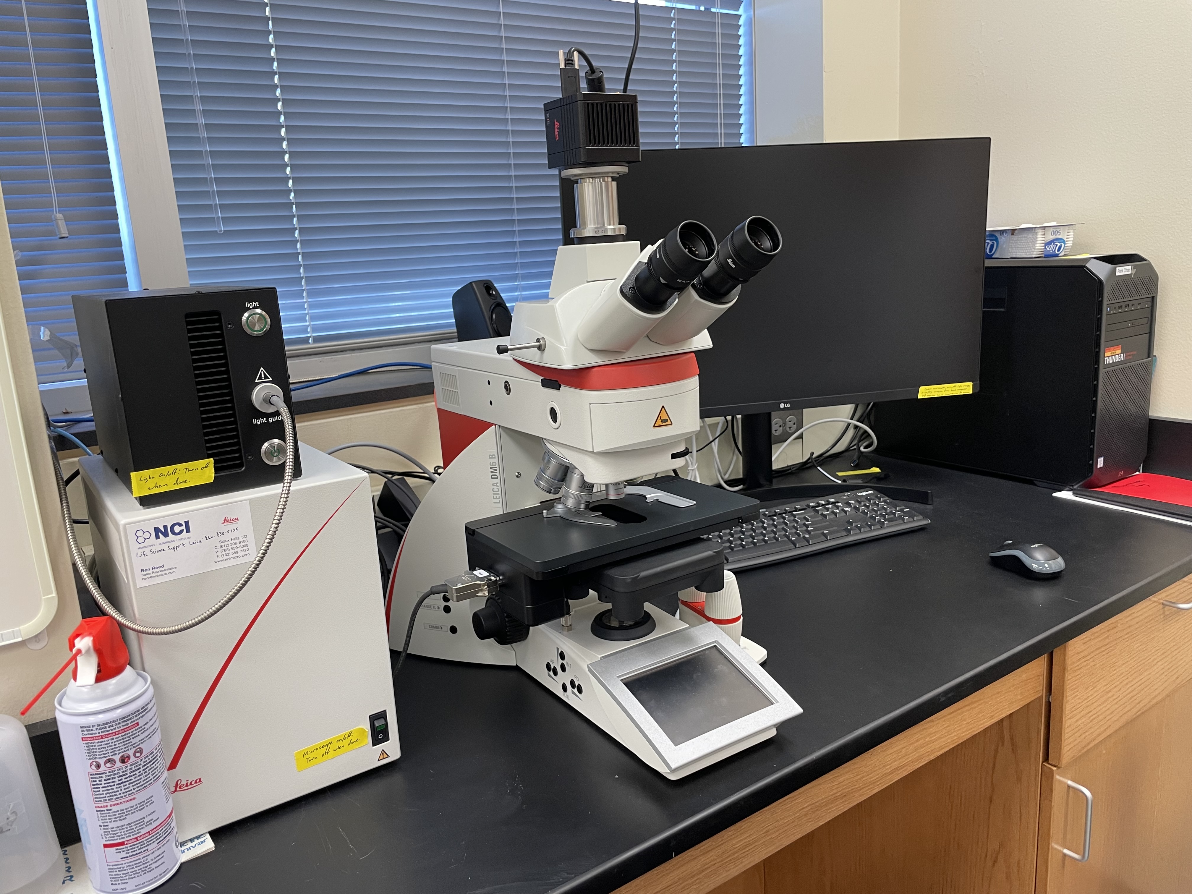
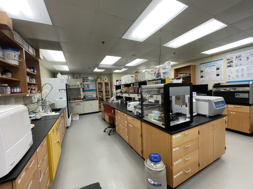
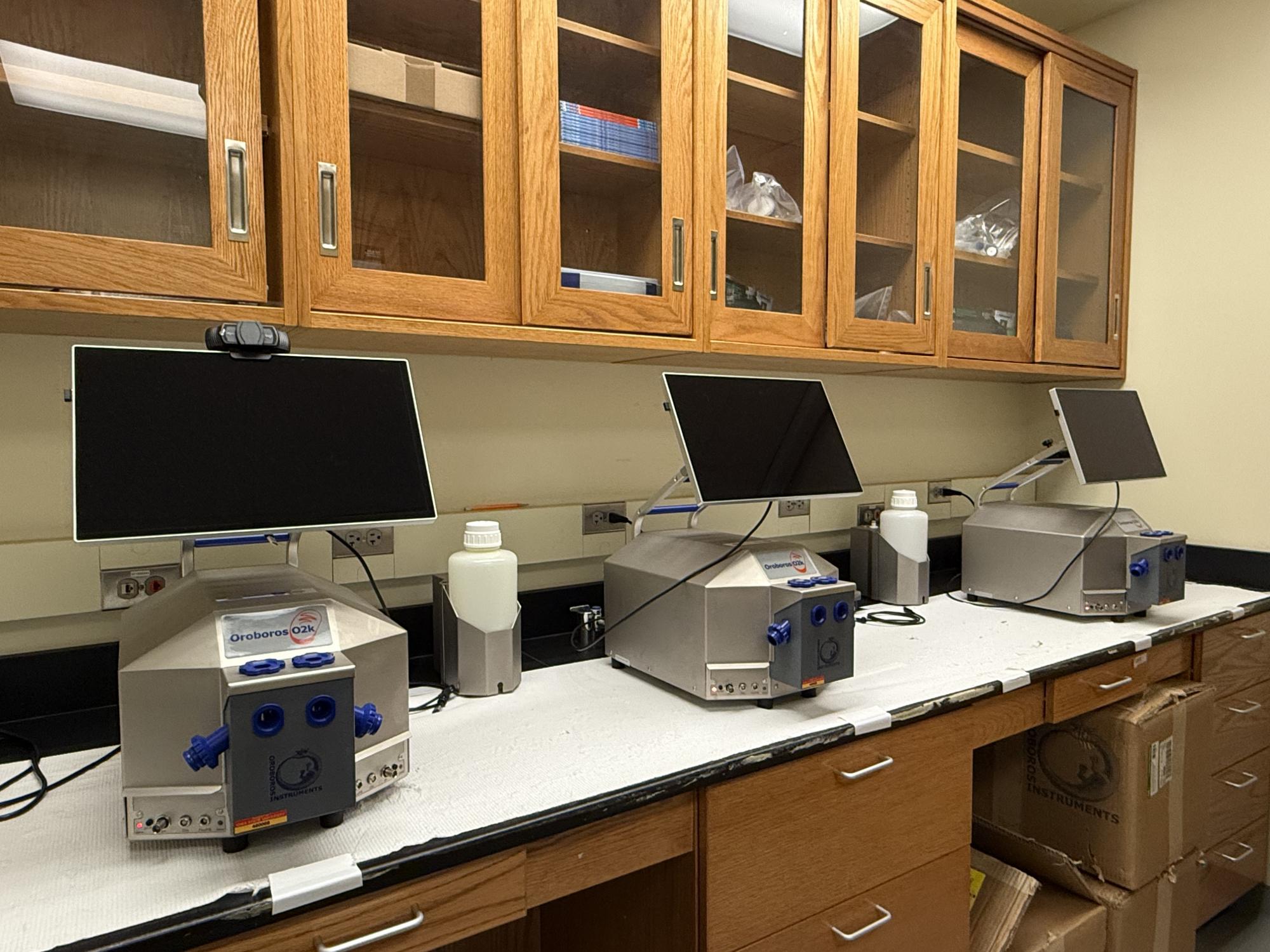
People & possibilities
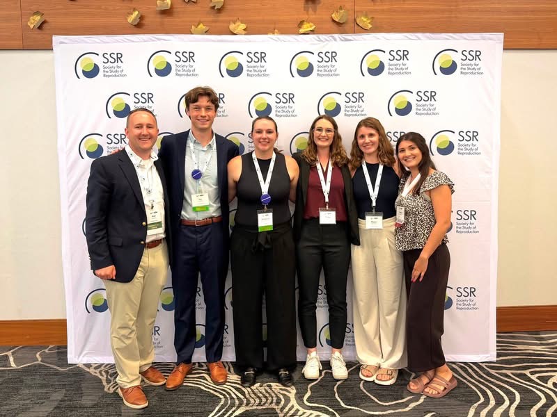
We are researchers, collaborators, and mentors committed to careful science and meaningful impact. Graduate students and collaborators are invited to start a conversation.
Meet the teamTomorrow’s discoveries are being unearthed today.— Kerns Lab
From the lab